The Anchor Atlas: A New Map of the Brainstem
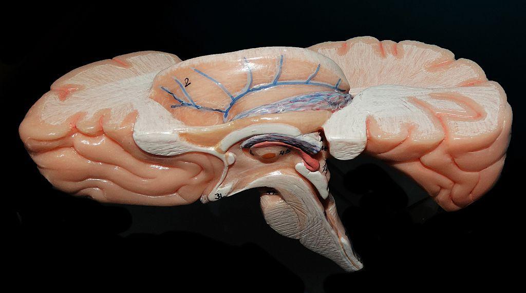
Researchers at the Sudha Gopalakrishnan Brain Centre (SGBC) at the Indian Institute of Technology, Madras, have created a digital map – the Anchor Atlas – that captures the human brainstem in three dimensions at cellular resolution. Using over 500 tissue sections from foetal to adult brains, the atlas links whole‑brain MRI data with microscopic anatomy, allowing scientists to move seamlessly between the macro‑scale structure and individual neurons.
Why the Brainstem Matters
The brainstem, though small, controls breathing, heartbeat, sleep and movement. Its densely packed architecture has long hindered detailed mapping, leaving clinicians with limited information when diagnosing disorders such as Parkinson’s or stroke. The Atlas identifies more than 200 clusters of brain cells and nerve pathways, using eight chemical markers to differentiate cell types.
Bridging Two Worlds of Imaging
Neuropathologists traditionally examine a brain en masse under the microscope, then sift through a few isolated sections. Modern MRI offers a bird’s‑eye view but lacks cellular detail, while microscopes reveal single cells in isolation. Anchor merges these two perspectives, providing an online viewer that lets users zoom from the whole brainstem down to individual neurons while maintaining their precise spatial relationships.
Future Implications and Cross‑Timeline Insights
The Atlas is freely available at https://anchor.humanbrain.in/, inviting researchers worldwide to explore its depths. By comparing healthy and diseased maps, scientists could uncover how conditions like Alzheimer’s, Parkinson’s and even COVID‑19 alter brain tissue cell‑by‑cell. In alternate future timelines, better understanding of the brainstem may lead to targeted therapies that preserve injured tissue, reduce perioperative complications in neurosurgery, and pave the way for personalized neurorehabilitation protocols.
Beyond Anatomy – A Platform for Innovation
Anchor’s approach, built from high‑resolution microscope images rather than costly molecular techniques, offers a scalable and affordable way to chart brain tissue. The SGBC now plans to image over 100 whole human brains across ages and diseases, creating a reference library that may reveal how disease reshapes the brain at the cellular level. While the Atlas itself does not diagnose, its capacity to answer new questions could shift the trajectory of neurological research and care across timelines that we are only beginning to imagine.
Image sources: Universal Images Group via Getty Images; SGBC; Getty Images.